Persistent leg pain, numbness or weakness? Arrange a consultation for comprehensive evaluation and management options.

Lumbar radiculopathy (often called sciatica) is nerve pain that starts in the lower back and travels down the leg, usually due to a pinched or irritated nerve in the lumbar spine. Many people improve with time and conservative care, but some benefit from targeted injections or surgery, depending on symptoms and nerve pressure.
Shooting or burning pain from the lower back into the buttock, thigh, calf or foot (often worse than the back pain itself).
Numbness, tingling or “pins and needles” in the leg or foot following a specific nerve pattern.
Weakness in the leg or foot, or feeling the leg might “give way”.
Pain that worsens with sitting, coughing, sneezing or bending, and may ease when lying flat or walking.
Red‑flag symptoms (e.g. difficulty passing urine, loss of bowel control, numbness around the groin, sudden severe weakness) need urgent medical assessment.
Lumbar disc herniation: a slipped or bulging disc pressing on a nerve root (most common).
Spinal stenosis: narrowing of the spinal canal or nerve exit holes.
Degenerative changes: age‑related wear, bone spurs and joint thickening around the nerves.
Less commonly, cysts, tumours, fractures or infection affecting the nerve root.
Diagnosis usually combines a careful history, examination and, when needed, imaging.
Clinical assessment: testing strength, reflexes, sensation and nerve tension (e.g. straight leg raise) to map which nerve is affected.
Imaging: MRI is the main scan to show disc herniation, stenosis or other structural compression; CT may be used in some cases.
Nerve tests: nerve conduction studies or electromyography (EMG) may be used if the diagnosis is unclear or symptoms persist.


Most people start with non‑surgical (conservative) care, especially if pain and weakness are manageable and there are no red‑flag signs.
Activity modification and education: staying as active as possible, avoiding prolonged bed rest, guidance on posture, lifting and pacing.
Medications: simple pain relief, anti‑inflammatories (if suitable), and sometimes short courses of neuropathic pain medicines or muscle relaxants.
Physiotherapy: targeted exercises to improve mobility, core strength and nerve mobility, plus manual therapy where appropriate.
Epidural or nerve root injections: image‑guided corticosteroid injections around the affected nerve to reduce inflammation and pain, often used when symptoms persist despite basic measures.
Conservative treatment is often trialled for several weeks, provided symptoms are stable and there is no significant or progressive neurological deficit.
Surgery is usually considered when pain remains severe despite adequate conservative care, when there is progressive weakness, or when serious nerve compression is present.
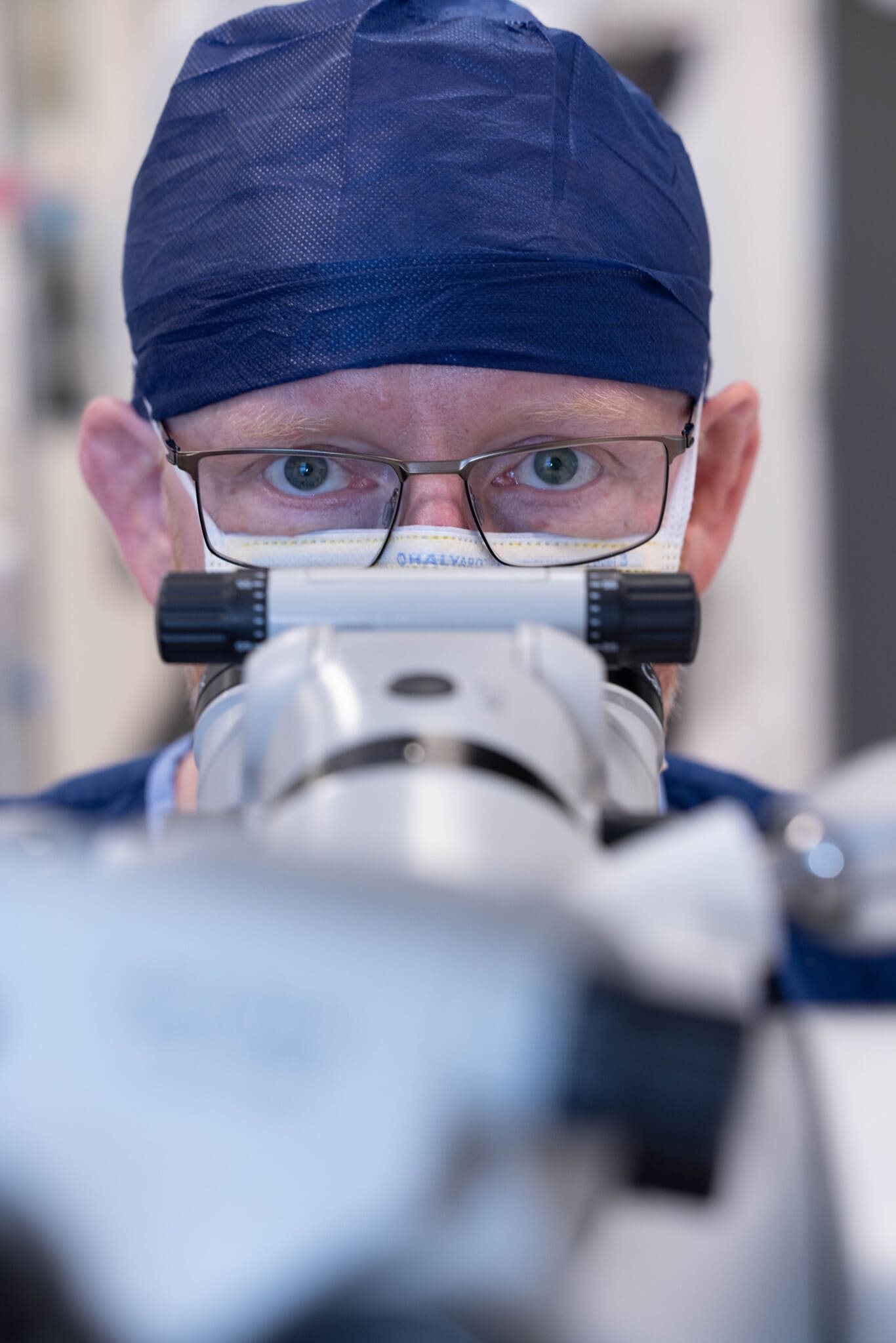
Lumbar microdiscectomy: removal of the part of the disc pressing on the nerve through a small incision, often using microscopic or endoscopic techniques.
Decompression (laminotomy/foraminotomy): removing bone or ligament to widen the nerve space, particularly in spinal stenosis.
In selected cases, stabilisation or fusion may be recommended if there is significant instability or deformity as well as nerve compression.
Evidence suggests surgery can provide faster pain relief for suitable patients, while long‑term outcomes may be similar to well‑managed conservative care.